Have any questions?
+44 1234 567 890
Projects
ARTURO - Augmented Reality (AR) in neurosurgery, development of an AI-based AR system for placing catheters in the brain for external ventricular drainage
The aim of the project and its innovation is the development of an AI-based augmented reality system for the placement of catheters in the brain for external ventricular drainage (EVD). In contrast to the predominantly freehand positioning of the catheter in the ventricle of the brain, the path of the catheter through the brain is virtually superimposed as a trajectory in the surgeon's field of vision with the help of AR data glasses or a smartphone and superimposed on the real patient anatomy. This system should enable surgeons to double the accuracy associated with the hit rate and thus significantly increase patient safety. The digitalization of surgical procedures using this form of computer assistance could therefore also benefit smaller hospitals on the periphery.
External ventricular drainage (EVD) is a life-saving measure to remove excess cerebrospinal fluid (CSF) from the ventricular system of the brain. This may be necessary if the ventricular system is blocked or if the brain is producing too much cerebrospinal fluid. Invasive EVD involves the use of drainage probes that are inserted through the skull into the ventricles and connected to a drainage device.
funded by: ![]()

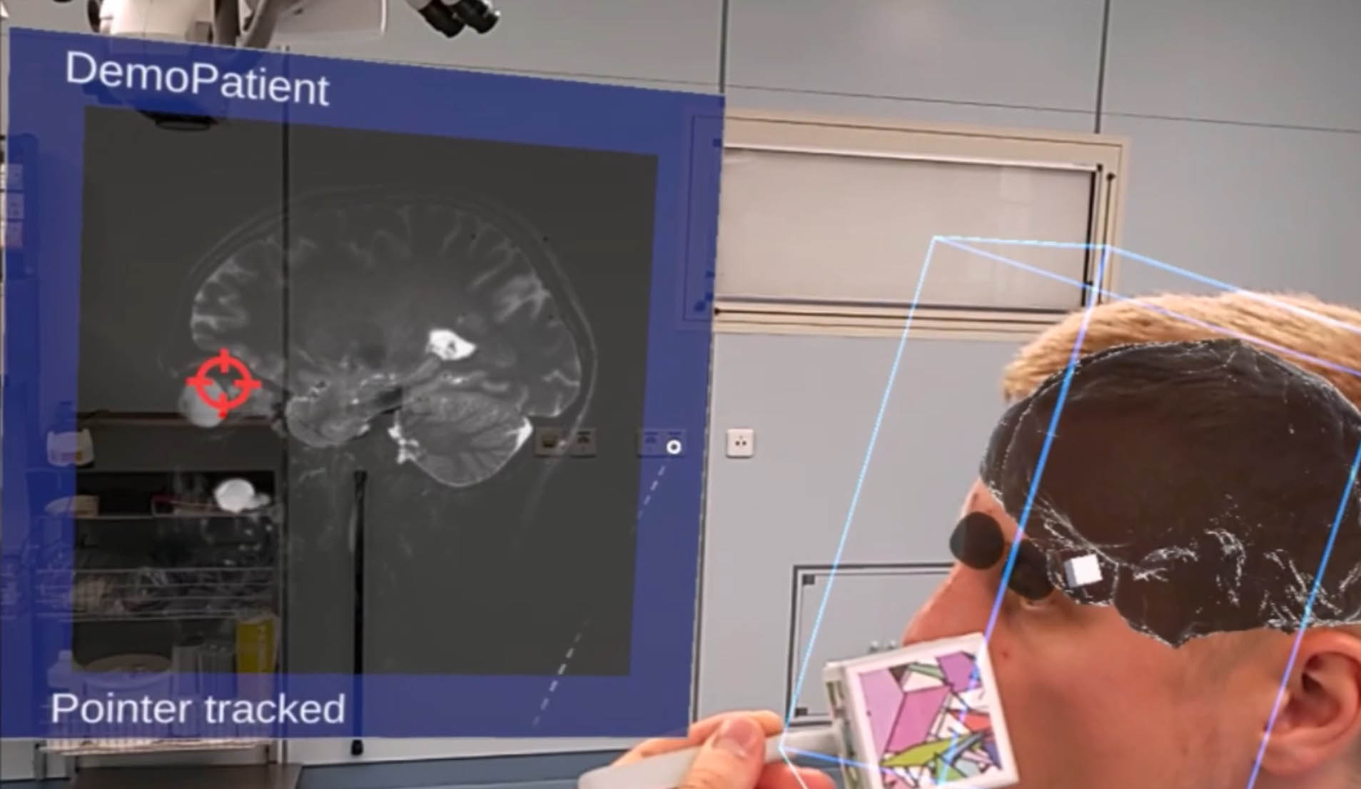
Development of a surgical AR navigation system
Chirurgische Navigationssysteme nehmen in der Hochpräzisionschirurgie einen hohen Stellenwert ein. Sie dienen als Orientierungshilfe und finden in der exakten intraoperativen Positionsverfolgung von chirurgischen Instrumenten Anwendung. Der intraoperative Abgleich von OP-Situs mit präoperativen Bilddatensätzen trägt dazu bei, Ziel- und Risikostrukturen besser identifizieren zu können und schlussendlich, Operationen sicherer durchzuführen. Unzweifelhaft bietet die Kopfchirurgie (MKG, HNO, Neurochirurgie) aufgrund des fixen knöchernen Rahmens von Gesichts- und Hirnschädel als reproduzierbare Referenz beste Voraussetzungen, dieses Konzept umzusetzen, besteht aber gerade hier die Herausforderung, auch im Bereich anderer chirurgischer Disziplinen diese Technologie aufzugreifen und als Instrument zu etablieren.
Surgical navigation systems play an important role in high-precision surgery. They serve as an orientation aid and are used for exact intraoperative position tracking of surgical instruments. The intraoperative comparison of the surgical site with preoperative image data sets helps to better identify target and risk structures and ultimately to perform operations more safely. Head surgery (maxillofacial, ENT, neurosurgery) undoubtedly offers the best conditions for implementing this concept due to the fixed bony frame of the facial and cranial skull as a reproducible reference, but the challenge here is to also take up this technology in other surgical disciplines and establish it as an instrument.
The aim of our navigation technology is to make navigation available as a reliable tool to many surgical sub-disciplines and users. We see the use of data glasses to superimpose augmented reality as a realistic solution for achieving this goal. And although the concept of augmented reality appears to be omnipresent and usable, the development of data glasses as a navigation aid and the interaction with surgical instruments and preoperative image data that this aims to achieve represents a considerable leap forward in development and a novelty at the same time.
funded by Dr. Hubertus von Grünberg Stiftung

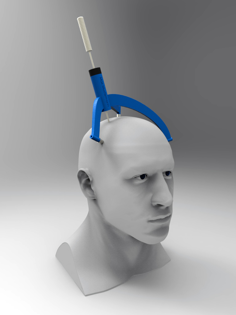
Development of a patient-specific system for deep brain stimulation
The aim of the project and its innovation is the development of a patient-specific stereotactic device for deep brain stimulation, which already implements all spatial coordinates in semi-automatic production and can be used directly for surgery. For the first time, this should eliminate time-consuming adjustments and adaptations for the respective patient, as required by all previously used procedures, and help to eliminate the potential for inaccuracies and incorrect settings.
Deep brain stimulation is a treatment technology that is used to treat movement disorders such as Parkinson's disease to minimize uncontrolled tremors.
funded by: 
Development of a patient-specific system for deep brain stimulation
The aim of the project and its innovation is the development of a patient-specific stereotactic device for deep brain stimulation, which already implements all spatial coordinates in semi-automatic production and can be used directly for surgery. For the first time, this should eliminate time-consuming adjustments and adaptations for the respective patient, as required by all previously used procedures, and help to eliminate the potential for inaccuracies and incorrect settings.
Deep brain stimulation is a treatment technology that is used to treat movement disorders such as Parkinson's disease to minimize uncontrolled tremors.
funded by: ![]()

Development of a computer-assisted system to support the planning and execution of tumor resections
The aim of the project and innovation is the development of a system for augmented intraoperative information display in neurosurgery. The main area of application will be the removal of tumorous brain tissue or tumor resection (e.g. glioblastoma). The main target parameters are a significant reduction in operating time and an increase in precision in order to avoid intraoperative trauma. The system is intended to achieve this by avoiding the surgeon's constant change of focus between the situs and the surgical monitor. Components of the project are the image data fusion of different modalities, intraoperative image acquisition and the development of a 3D printing-based test scenario.
funded by:
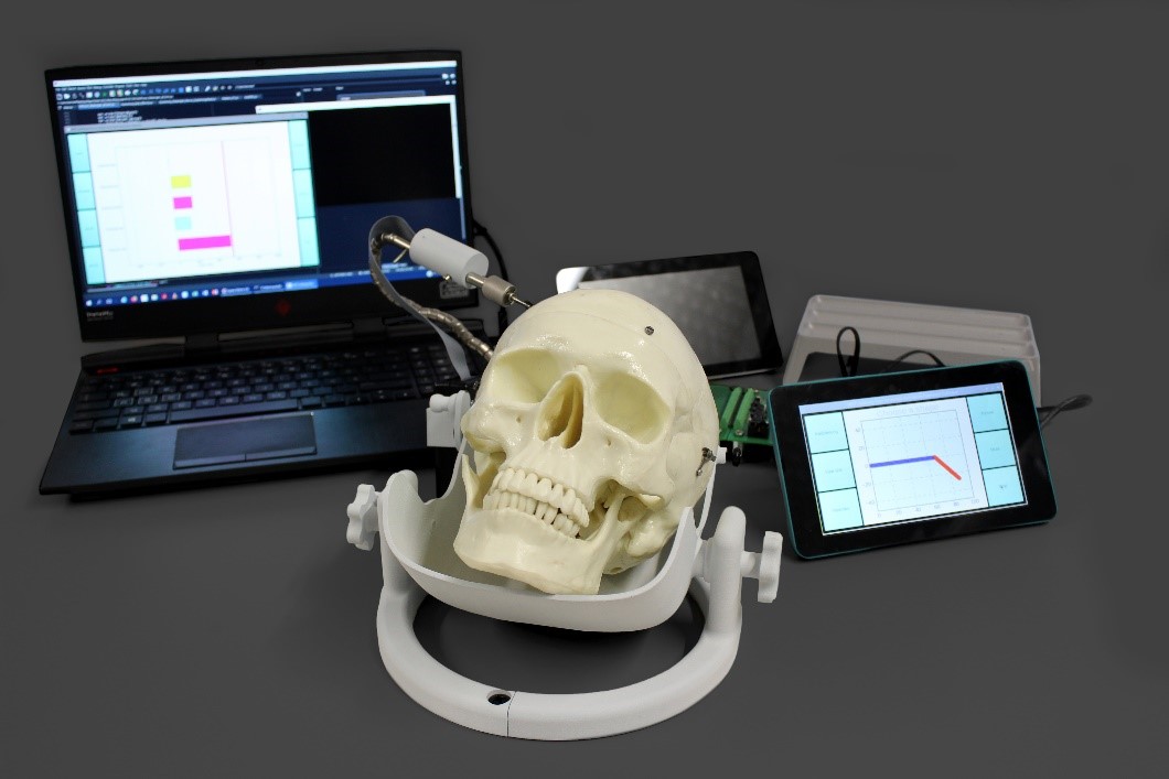
Development of an intelligent brain spatula to determine the intraoperative load on the brain parenchyma during neurosurgical procedures
During neurosurgical procedures on the open brain, so-called brain spatulas are used to separate the highly sensitive brain tissue (parenchyma) and reach deeper target regions inside the brain. The aim of the project is to develop a new type of brain spatula with an integrated sensor function to record the mechanical force applied to the brain tissue and to send a signal in the event of impending brain tissue damage. The data collected by the sensor system will be available to the operating neurosurgeon both intra-operatively (monitoring) and post-operatively. The latter is to be used as a data evaluation unit for quality assurance and risk minimization for future neurosurgical interventions.
funded by:
Development of planning software for deep brain stimulation - probabilistic tractography
The aim of the planned THSpro research project is to develop a software tool for target point planning for deep brain stimulation. The neuronal fiber tracts in the brain are reconstructed from measurement data from diffusion-weighted magnetic resonance imaging using probabilistic tractography. This neurological-functional data is used to determine the target point for deep brain stimulation. It will improve the accuracy of target point determination and thus directly serve the patient's well-being. The process-controlled procedures of the planning process are optimized to save time and thus costs. Training and operating assistance is efficiently realized with an interactive video interface. A specially designed VSEO strategy will help experts and those affected to obtain comprehensive information.
Machine learning methods are used to shorten the time-consuming calculations of probabilistic tractography. This means a more time-efficient planning process and saves costs.
funded by:

Patient-specific 3D-printed helmets for children
Infants can have a deformed head due to various circumstances. This can occur, for example, prenatally due to constriction in the uterus, perinatally due to trauma during birth, such as breech presentation, and postnatally due to functional or anatomical constraints or a permanent supine position.
The helmet orthosis utilizes the growth of the head to correct its shape. It lies against the prominent areas of the head, so growth is suppressed here during the treatment period. It leaves space in the flattened areas and allows growth into the ideal shape.
funded by:

Development of a system for planning and manufacturing patient-specific cranial implants using additive manufacturing processes and machine learning techniques
The aim of the project is to develop a process chain for the production of patient-specific cranial implants using 3D printing systems, with which cranial implants made of various materials such as PEEK / PEKK, bioceramics and titanium can be produced individually for each patient. For CT-based preoperative planning of the patient-specific cranial implant, software is currently used that requires several hours to segment the relevant anatomical structures and create the CAD model of the implant. During the implant planning process, the interaction between the surgeon and the engineer is currently not optimal. Another project goal is therefore to develop a cloud-based planning software with artificial intelligence to segment the relevant anatomical structures within the CT images and automatically create different design versions of the implant. The entire process should enable implant delivery within 48 hours instead of the previous 2-6 weeks.
funded by:

Patient-specific AR and 3D-printed models for planning complex operations and implant placement
As part of precise bone screw fixation or bone repositioning, drilling and sawing templates can be produced for spinal and pelvic surgery as well as for oral and maxillofacial surgery.
In addition, patient-specific AR or 3D print models of the patient's anatomy enable the safe planning of complex operations.
Patient-specific 3D-printed vascular models
When treating life-threatening aneurysms of the main artery, the aorta, the coordinates of the vessel branches are transferred from the X-ray images to the vascular implant during the operation, which is very time-consuming. Using 3D-printed patient-specific vascular models as templates, vascular branches can be marked on the vascular implant.
This allows the vascular implant to be placed in the patient with high precision and 90 minutes faster.
MUSOK Development of a musician-specific earmold with integrated filter for manual or individual adjustment of the filter effect directly on the ear
The aim of the project is to develop a passive, musician-specific and manually adjustable hearing protector and the associated digital process chain for creating it. This hearing protection should be an earmold that changes the filter effect by manually turning it. In addition to the precise and comfortable fit of the hearing protection, it is important to simplify the creation of the filter systems using a digital process chain. The development of such a system or the further development of existing systems has the potential to significantly improve the quality and integration/acceptance of such hearing protection systems. Accordingly, the occurrence of early onset hearing loss or acute disorders of the auditory system in musicians can be prevented as far as possible.
funded by:

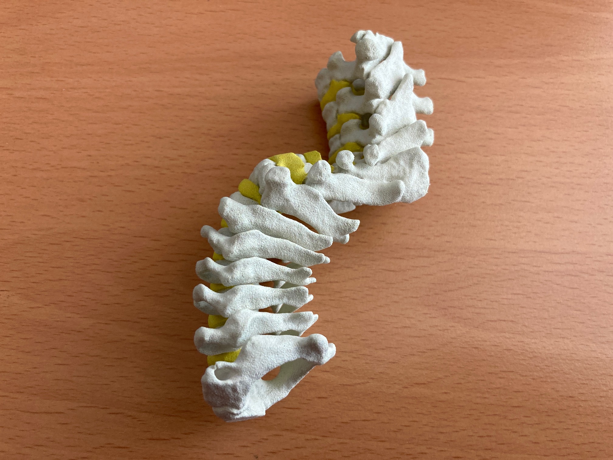
Development of shape memory implants
The development focus is on transferring the concepts for shape memory implants to the development of a shape memory cage for an anatomically adapted disc replacement. With the help of the thermal shape memory effect, a cage-like disc replacement is to be developed that can be implanted without damaging the tissue and unfolds and supports itself between the vertebral bodies when activated by body heat. The innovative approach is the anatomical adaptation of the cage geometry to the intervertebral space. In the area of the cervical spine, an existing concept for spinal implants made of shape memory alloys is to be optimized in order to protect critical nerve structures and the body's own tissue.
funded by: 
Fossa Cranii - "Individualized 3D printing cranioplasty after unilateral trephination of the posterior fossa to avoid cerebrospinal fluid fistulas and postoperative scar pain
Surgical reconstruction of the skull defect (cranioplasty) is routinely performed after an open neurosurgical operation, such as a tumor resection. In the case of larger defects, artificial materials are used to restore the stability of the skull. In the case of smaller defects, the original bone fragments are reimplanted as standard, although this can be problematic in the case of extended trepanation. When removing acoustic neuromas in the posterior fossa, the initial craniotomy usually has to be extended piece by piece to ensure optimal resection in this complicated anatomical region without damaging surrounding tissue. This results in a defect after reimplantation of the bone fragment, known as a trephination defect, which increases the postoperative complication rate in the form of ligament fistulas and subsequent scar pain. This results in longer hospital stays and possibly repeated surgical interventions. To date, there are no surgical solutions on the medical device market to tackle this problem with the help of patient-specific defect covers. This is precisely where the proposed project should come in.
The aim of our navigation technology is to make navigation available as a reliable tool to many surgical sub-disciplines and users. We see the use of data goggles to superimpose augmented reality as a realistic solution for achieving this goal. And although the concept of augmented reality appears to be omnipresent and usable, the development of data goggles as a navigation aid and the interaction with surgical instruments and preoperative image data that this aims to achieve represents a considerable leap forward in development and a novelty at the same time.
- Preoperative solution approach
During surgical planning, access to the skull is to be developed with the help of CT and MR images. A milling template is produced accordingly, which the surgeon mills along during the operation. The surgical field is thus defined and a suitable implant can be produced preoperatively.
- Intraoperative solution approach
During the operation, the trephination defect is scanned using a 3D scanner and the data is then evaluated. From this, a custom-fit implant can be produced. The aim is to insert the implant in the same operation (in real time).
funded by:

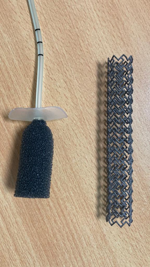
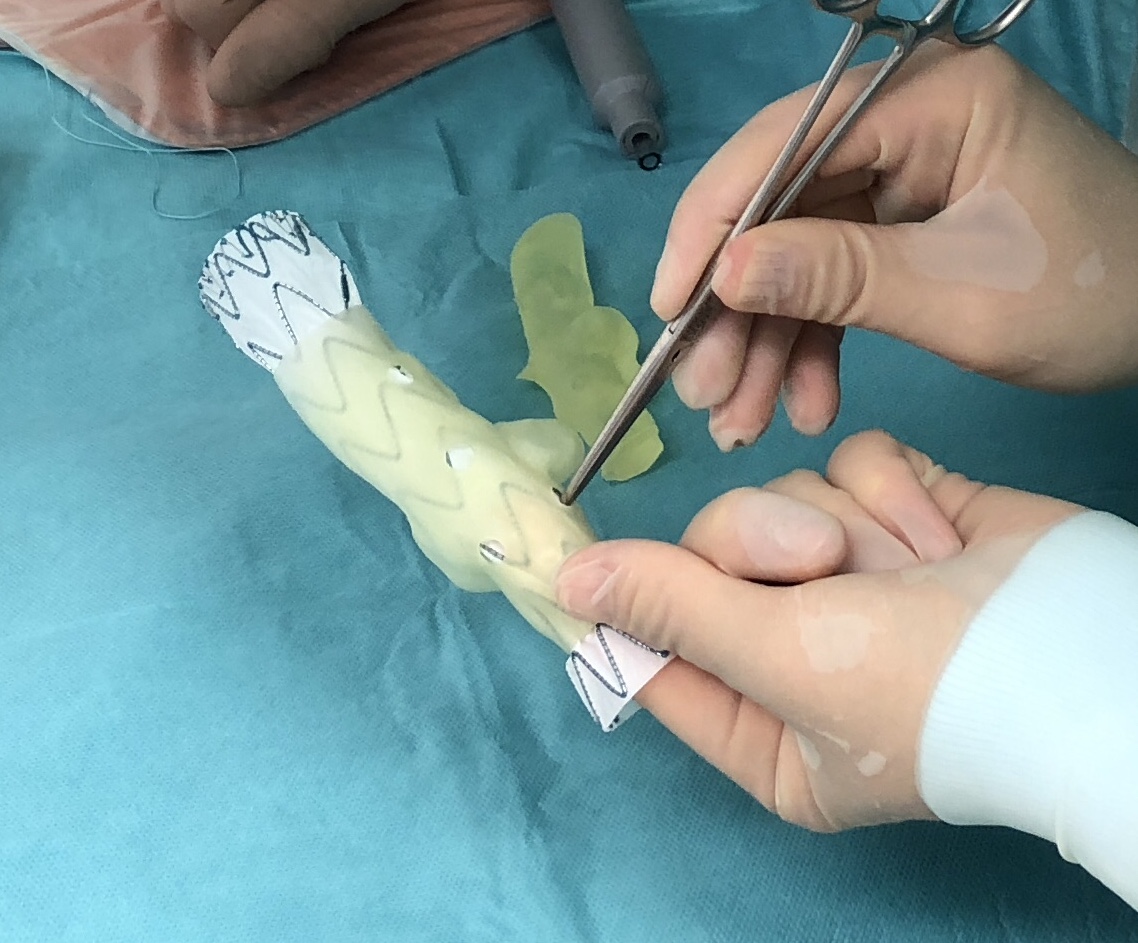
SpongeEx - method for performing stent-over-sponge therapy using a patient-specific system for placing a sponge in the oesophagus
In very severe cases of esophageal complications, a stent is also implanted over the polyurethane sponge in the esophagus. These stents are fine scaffolds made of nitinol or polyamides to keep anatomical structures open and are implanted using special delivery systems.
The sponge is changed every two to three days in a complex procedure, whereby the stent is first removed orally and then the sponge is pulled out of the esophagus using an endoscope. Both components are implanted again one after the other. Until now, there has been no way to remove and reinsert the sponge from the esophagus independently of the stent. This is precisely where the planned project comes in.
The aim of the project is to develop a new method for changing the sponge in the "stent-over-sponge" therapy. The aim is not only to significantly improve patients' quality of life and chances of survival, but also to minimize the time required for treatment and thus the costs for the hospital.
funded by: